Chest wall tumours
Chest wall tumours are tumours occurring in the walls of the chest cavity. They are characterized by abnormal cell growth that can be cancerous or non-cancerous. However, around 60% of all chest wall tumours are cancerous.
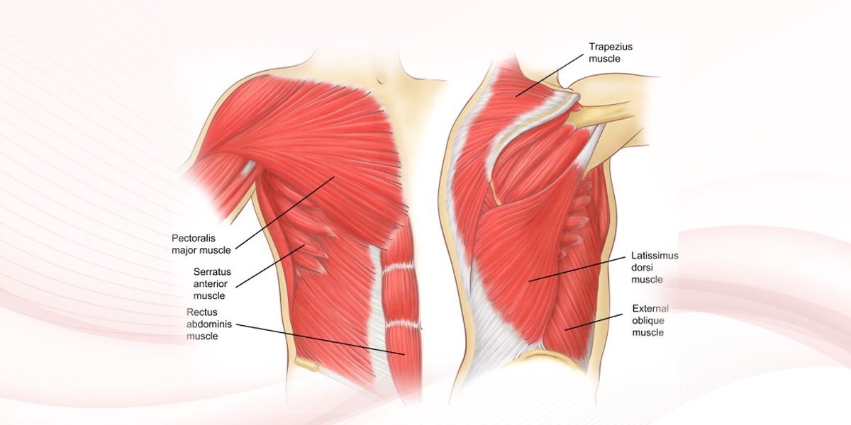
Your chest walls are made of soft tissues, bone, and cartilage. Chest wall tumours can form in any of these areas. In most cases, tumours are formed in the bone (especially the rib cage) or the cartilage.
There are two major types of chest wall tumours – primary and secondary. Primary chest wall tumours begin in your chest walls and keep growing if rendered untreated. Secondary chest wall tumours begin somewhere else and spread to the chest wall.
How Common Are Chest Wall Tumours?
Less than 1 in 50 people are affected by primary chest wall tumours. The most common non-cancerous chest wall tumours are chondromas (slow-growing tumours made up of cartilage forming in bones or soft tissues) and osteochondromas (a bone and cartilage overgrowth occurring at the bone’s end near the growth plate). The most common cancerous chest wall tumours are sarcomas (cancers including fat, cartilage, muscle, blood vessels, fibrous tissue, or other connective tissue originating from the bone or soft tissue).
In most cases, the chest wall tumours found in children are primary. On the other hand, secondary chest wall tumours are common in adults.
Symptoms of Chest Wall Tumor
- Swelling in the affected region
- A localized mass
- Chest pain
- Muscle atrophy (breakdown)
- A malignant (cancerous) chest wall tumour will have similar symptoms, chest expansion, and/or impaired chest movement.
Here are a few more signs that should prompt you to seek medical help regarding chest wall tumours:
- Pain, tingling, or weakness in the arm or shoulder, especially while raising your arms
- Unusual appearance of the chest due to chest wall malfunctioning
- Reduced tolerance for exercising
- Increased heart rate
- Fatigue
- Shortness of breath
Diagnosis of Chest Wall Tumor
While there are no clear causes for chest wall tumours, factors like diet, lifestyle choices, and heredity are known to have increased the risk of the tumours.
Chest Wall Tumours: Diagnosis And Treatment
Chest wall cancers can be diagnosed via biopsies and imaging tests (X-ray, CAT Scan, PET scan, MRI, etc.). Healthcare providers may run these tests if you report the right symptoms and they sense the need for a thorough diagnosis. At times, doctors may also diagnose chest wall tumours using imaging tests done for other purposes (incidental diagnosis).
Generally, imaging tests confirm the presence of chest wall tumours. Our Chest Specialist may suggest a biopsy if the tumours are not found through these tests despite the symptoms.
Once diagnosed, the treatment for a chest wall tumour generally includes the following:
Surgery (resection) for removing the tumour
Reconstructive surgery for repairing the damage your tumour caused to the chest wall Radiation therapy and chemotherapy, if the tumour is cancerous Our doctor will give you the most suitable timing and sequence for different treatments. For example, they may recommend chemotherapy before surgery to reduce the tumour’s size, making its removal easy.
Once your chest wall tumour treatment is completed, the doctor will explain a follow-up plan to you and your family members, letting you know how often you need to visit for follow-ups.

Dr Shashikiran N J
MBBS, MS, MCh
Lead Consultant Minimally Invasive Thoracic surgery
Apollo Hospitals Hyderabad